Harvard University scientists are slicing mouse brain segments a mere 29.4 nanometers thick, imaging them under elaborate microscopes and reassembling the images into a representation of the brain. This arduous process will help researchers construct a connectome - a comprehensive map of neural connections in the brain demonstrating how memories, traits and skills are stored.
The field of connectomics has grown largely because of the National Institute of Health, which, in September, granted $40 million toward such research at top universities. The ultimate goal is to create a human connectome that illuminates the connections between 100 billion neurons. A human connectome would provide intricate insight about brain function and mental illness. Surgeons also would be capable of delicately slicing areas of white matter, a component of the central nervous system, crucial to treating illnesses such as epilepsy.
The effort needed to assemble a human connectome, however, is overwhelming. Although some scientists believe in the value of developing a connectome, which essentially produces a momentary image much like a photograph of the brain, others argue that this will hinder researchers from observing dynamic brain responses.
-compiled by Sasha Gitomirski
Read More

U.Va. students and physicians team up for another year of free kidney screenings
By Merrill Hart | November 5, 2024Saturday’s screening marked the first by KDSAP at Portico Church, near IX Art Park.
U.Va. Health sees a spike in COVID-19 cases
By Emily Horn | September 4, 2023U.Va. Health ended their vaccination requirements in August, and masks were made optional in most hospital areas in early April.
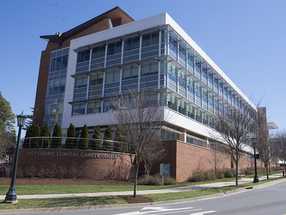
U.Va. Cancer Center harnesses $12 million grant to support opportunities for undergraduates, ongoing research and new center
By Ellen Wu | January 12, 2023The grant provides an opportunity to establish the Center for Systems Analysis of Stress-Adapted Cancer Organelles, which seeks to find the cause of cancer in individual organelles.