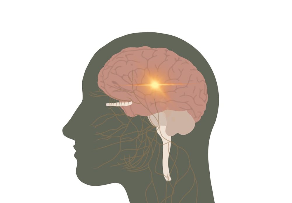
About 50 million people globally are affected by epilepsy, making it one of the most common neurological diseases. But if properly diagnosed and treated, 70 percent of those impacted could control their recurring seizures. To aid in this work, a team of University researchers — funded by the U.Va. Brain Institute — created a new approach for diagnosing epilepsy, using an enhanced form of positron-emission tomography, which can better recognize areas of the brain where the chance of epilepsy is higher.
PET is a diagnostic tool for epileptic patients that measures glucose use in the brain. A normal PET scan identifies brain regions that use significantly less sugar than other areas as bad epilepsy spots by using a dye containing radioactive tracers injected intravenously to show the activity in affected areas. Although PET is generally successful in identifying epilepsy-affected areas, its accuracy is low and it cannot always identify specific targets well.
U.Va. Health neurologist Mark Quigg, radiology and medical imaging Assist. Prof. Bijoy Kundu and Class of 2020 alumnus Vikram Seshadri launched a small pilot study using seven participants to test out a new technique to improve how diagnoses are made. Their goal was to refine the method of glucose mapping for more accuracy and better resolution.
“Normally in a PET scan when we're taking a picture of glucose use in the brain we use a radioactive tracer,” Quigg said. “What [Kundu] has invented is a process of a dynamic PET.”
In the dynamic PET scan, a tracer is injected into the bloodstream while the patient is already in the scanner. The scanner then takes pictures like a stop motion animation movie. The result is a time map of glucose uptake and glucose use in relation to brain region. The time map is used to identify points in time that yield the most difference among the functional places in the brain, or areas with most glucose uptake, and the nonfunctional places in the brain, or areas with least uptake.
The key difference between the former way of imaging and the dynamic method lies in the way the images are taken. Instead of producing a single snapshot of the brain, the dynamic PET captures a series of snapshots that then require mathematical modeling for image analysis.
“We are creating four dimensional images,” Kundu said. “Typical images are three dimensional x, y, z MR images.”
These dynamic series snapshots provide more information than a singular image and allow health professionals to see the stark difference between functional and nonfunctional brain regions when comparing brain scans from different epilepsy patients. Some areas are classified as in a hypometabolic state, a condition in which metabolic rate is strongly suppressed and normal activities are suspended.
On the scan, seizure locations are defined by blue clusters, which indicate low glucose regions, and the most blue clusters are typically found within the cerebrum — the part of the brain most associated with epilepsy.
In order to improve the results of the PET scan, however, the group also utilized an MRI — a structural imaging modality responsible for capturing the size of different parts of the brain. Combined use of the PET scan and MRI technology allows the researchers to align respective images in an overlay, which displays areas of high and low activity and enables the researchers to easily identify where the blue regions are located specifically, such as in the temporal lobe or frontal region.
This new technique poses a huge advantage because the results are more quantitative and rigid, which allows for clearer data and less subjectivity compared to the previous PET scan method.
Quigg believes that the findings from this research are very promising for new treatment plans. The new diagnostic approach is completely non-invasive and enables more accurate diagnoses.
“We can avoid going on to require invasive methods of determining where the bad spot is, which scares some people off,” Quigg said. “[Invasive methods] prematurely takes [people] out of surgical consideration because in some patients we have to implant electrodes deep in the brain.”
With a more effective diagnostic tool, the number of candidates eligible for epilepsy surgery will increase. Easier access to epilepsy surgery will provide patients with many benefits such as freedom from seizures, reduction in antiepileptic medication, and eradicating their disability.
Now, the research group is working towards experimenting with a bigger group of patients to validate the findings of the original data.
“The next step is to try to seek external funding for a larger grant, typically not just involving U.Va. but a group of like minded investigators,” Quigg said.
Read More

U.Va. Health opens new clinic in Pantops to expand patient access to plastic surgery
By Vrinda Vashisht | June 6, 2026U.Va. Health Plastic Surgery Pantops opened early May, moving aesthetic plastic surgery services from the University Hospital Grounds to a new clinic in Albemarle County.

U.Va. researcher finds potential way to treat Down Syndrome
By Lauren Seeliger | October 27, 2025Brandebura was hired by the University to start in her own lab — the Brandeura lab — and she will be continuing her work to study PTN.

STEM organizations host Neuroscience Outreach Fair for local K-12 students
By Lucia Gambacini | February 24, 2025The organizations invited elementary through high school students from across Albemarle and Fluvanna Counties to participate in a variety of neurology-related activities.